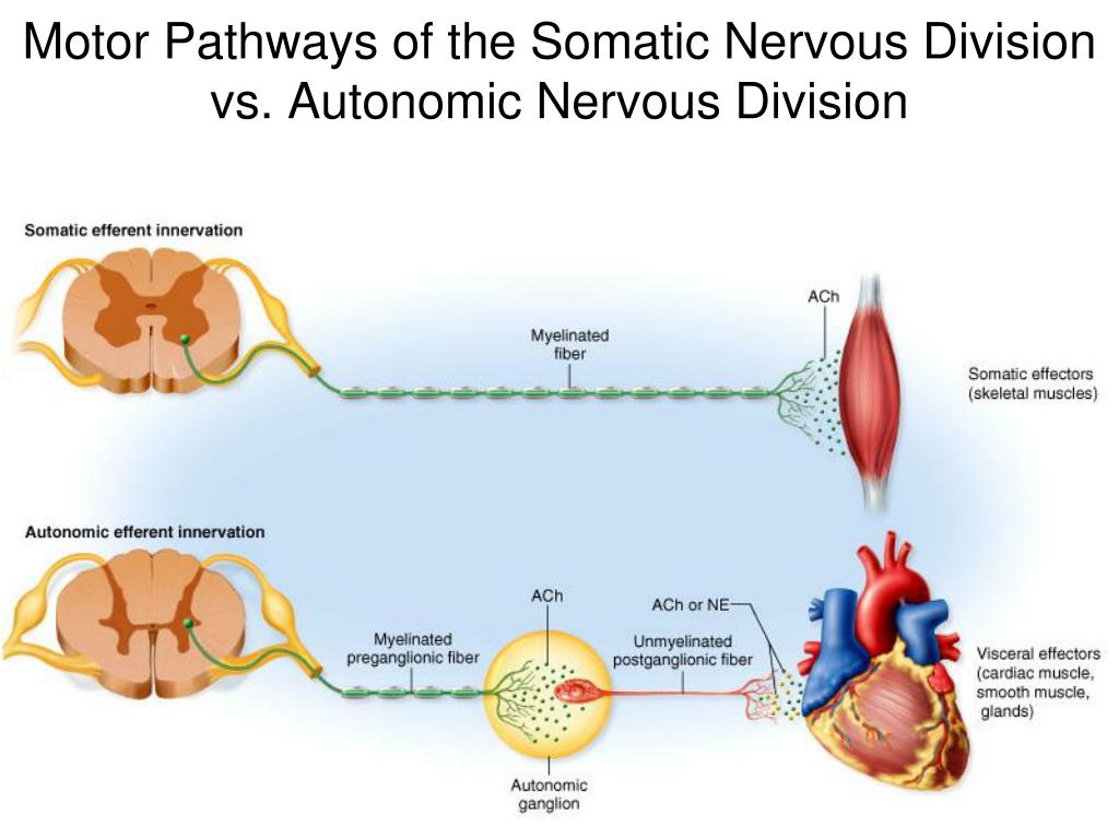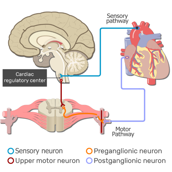
The spinal cord white matter consists of dorsal posterior columns, lateral columns, and ventral anterior columns. The spinal gray matter can also be divided into nuclei orusing a different nomenclatureintralaminar, named by Bror Rexed, with different functions that will be discussed later. Motor neurons send their axons out of the spinal cord via the ventral nerve root filaments. The central matter has a dorsal posterior horn that is involved mainly in sensory processing an intermediate zone that contains interneurons and certain specialized nuclei and a ventral horn, or anterior horn, that contains motor neurons. One branch conveys the sensory information from the periphery, and the other carries the information through the dorsal nerve root filaments into the dorsal aspect of the spinal cord. Sensory neurons in the dorsal root ganglia have axons that bifurcate. The spinal cord contains an "H" or butterfly-shaped central gray matter surrounded by ascending and descending white matter columns, also called funiculi. When looking at spinal cord anatomy, the cord is divided into gray matter, depicted here on the left, and surrounding white matter. A part of the body with fewer sensory and/or motor connections to the brain is represented to appear smaller. Because of the fine motor skills and sense nerves found in these particular parts of the body, they are represented as being larger on the homunculus. The resulting image is a grotesquely disfigured human with disproportionately huge hands, lips, and face in comparison to the rest of the body. These depictions are used to assist us for clinical localization of cortical function. The cortical maps depicted hereon the left, the sensory homunculus (homunculus meaning "little man" in Latin), and, on the right, the motor homunculuswere developed to assist us in understanding broadly that regions of the cortex correspond to regions in the body. This spatial orientation ensures that the point of origin of the signal is maintained all the way to the final point of termination of the pathway. Thus, the pathway is said to have somatotopic or spatial orientation. For example, information related to the lower part of the body travels in fibers located medially in the white matter of the spinal cord while those from the upper part of the body travel in fibers located at the lateral parts. Fibers in a pathway are arranged so that information from the lower part of the body travels in a particular pathway or part of the central nervous system that is different from information from the upper parts of the body. Somatotopic organization ensures that information from a specific area of the body is represented in a specific area of the cortex. Similarly, somatosensory association cortex in the parietal lobe receives inputs from the primary somatosensory cortex and is also important in higher order sensory processing.įunctional mapping and lesion studies have demonstrated that the primary motor and somatosensory cortices are what is described as somatotopically organized. These regions are involved in higher order motor planning and project to the primary motor cortex. There are several important areas of motor association cortex that lie just anterior to the primary motor cortex including the supplemental motor area in green and the premotor cortex in orange. The primary motor cortex in red is in the precentral gyrus while the primary sensory cortex is in the post-central gyrus. Remember that these areas are located on either side of the central sulcus, which divides the frontal lobe from the parietal lobe. Here, in slide 2, the primary sensory and motor areas are shown. At the end of this section, you should be able to: describe the anatomy and functions of the spinal cord including nuclei and laminae discuss spinal cord blood supply identify the site of origin, decussation, and levels of termination for the corticospinal tract and other motor pathways describe the autonomic nervous system, division, fibers, neurotransmitters, and regulation differentiate upper and lower motor neurons in the nervous system and describe common gait disorders in neurology. In this lecture, we will be looking at the organization of the motor system, specifically, the corticospinal tract and other motor pathways. Augustine for Health Sciences in San Diego, California. Annie Burke-Doe, a practicing physical therapist and an associate professor at the University of St. Welcome to Neuroanatomy in Physical Therapy.


Augustine for Health Sciences in San Diego, California Practicing physical therapist and associate professor at the University of St.


 0 kommentar(er)
